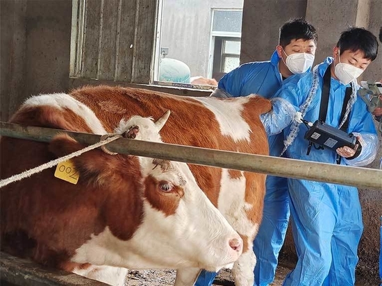
The S1 Handheld Veterinary Ultrasound Machine is a powerful tool that aids veterinary professionals in non-invasive diagnostics and monitoring, enhancing the precision and efficiency of veterinary care. It finds extensive application in pregnancy checks, liver, kidney, gastrointestinal, and bladder examinations for various animals, including cows, dogs, horses, sheep, ca...
Many cattle farmers do not know how to see whether the cow is pregnant or not, some cows are not pregnant but are raised as pregnant cows, which has a very big impact on the efficiency of the cattle farm. Here are several ways to judge whether a cow is pregnant or not, as well as the use of cattle ultrasound machine, I hope the majority of cattle farmers can help!
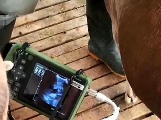
Among the many options available, the Dawei S1 veterinary ultrasound machine is undoubtedly a popular choice as it is ideally suited to the needs of cattle. The Dawei S1 veterinary ultrasound is suitable for a wide range of animals, not just cattle, and it can be used to examine the abdominal organs of a wide range of animals, providing accurate diagnosis and monitoring.
Esaote MyLab Alpha: This portable ultrasound machine from Esaote is known for its superior image quality and versatility. It is lightweight and easy to transport for a variety of veterinary applications. Mindray M8 Vet: Mindray M8 Vet is a compact portable ultrasound system designed for veterinary use. It features advanced imaging capabilities and a user-friendly interfa...
However, research has been conducted in recent years on the ultrasound imaging characteristics and examination areas of the gastrointestinal organs of cattle, goats, and other ruminant animals. Ultrasound has also been applied to the diagnosis of abomasal displacement (AD) and traumatic reticuloperitonitis.
One of the key features of the S0-VET is its portable design. The device is lightweight and easy to carry, making it a versatile tool for pregnancy examinations on the move. Long battery life: The S0-VET boasts impressive battery life, capable of running continuously for around 3 hours. This extended battery life gives users plenty of time for in-depth scanning session...
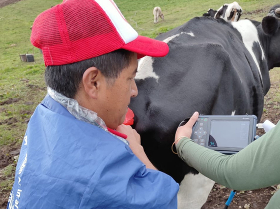
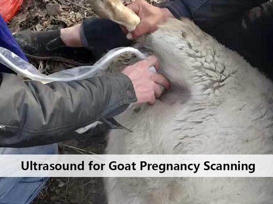
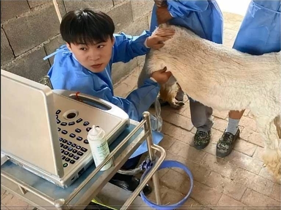
Advantages of using an ultrasound machine for pregnancy testing in sheep? The use of ultrasound in production in sheep, unlike in companion animals or horses, is an animal practice that is usually limited to reproductive management. Because of the small rectum and high fertility of sheep, which means that ultrasound imaging of ovaries sheep rarely have non-pregnant repro...
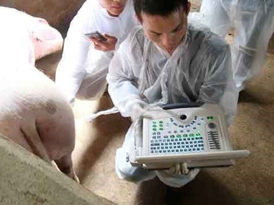
In modern livestock farming, ultrasound technology has become an indispensable tool for assessing animal health, monitoring disease and optimising farm management. For pig farming, choosing the right ultrasound technology can bring many benefits, helping to improve output efficiency, ensure animal well-being and promote sustainable livestock farming. The following Chines...
The use of swine ultrasound machine is receiving more and more attention in the daily production management of pig farms. Ultrasound for sows is also relatively common. Dawei Veterinary Medical summarize the the know-how of using swine ultrasound machine in practice to help you better use it. 1. The size and position of the sow’s uterus will change with the development ...
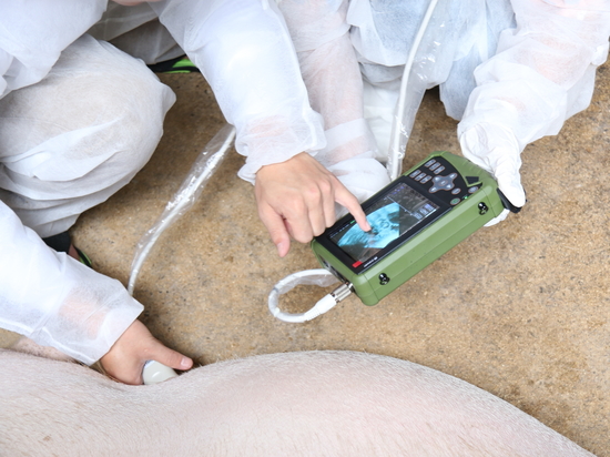
The steps to do ultrasound for sheep are roughly as follows: 1 Choose a suitable ultrasound device, preferably portable and capable of wireless transmission of images to a tablet or phone. 2 Prepare a clean scanning area, which can be a fixed barn or a mobile isolator. 3 Make sure the sheep's body is dry and shave off excess hair from the back and abdomen. 4 Apply an ap...
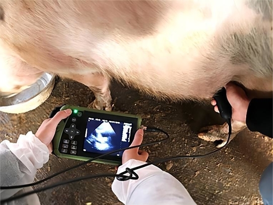
Ultrasound images of sow pregnancy The common veterinary ultrasound images generally show three colors: black, gray and white 1. Black: aqueous substances, such as amniotic fluid, blood, urine, pus, etc. 2. Gray: organ parenchyma. 3. white: denser objects, such as bones, teeth, organ fascia, etc. A: 22-30 days after mating is the best stage to observe the image, showing...