#Industry News
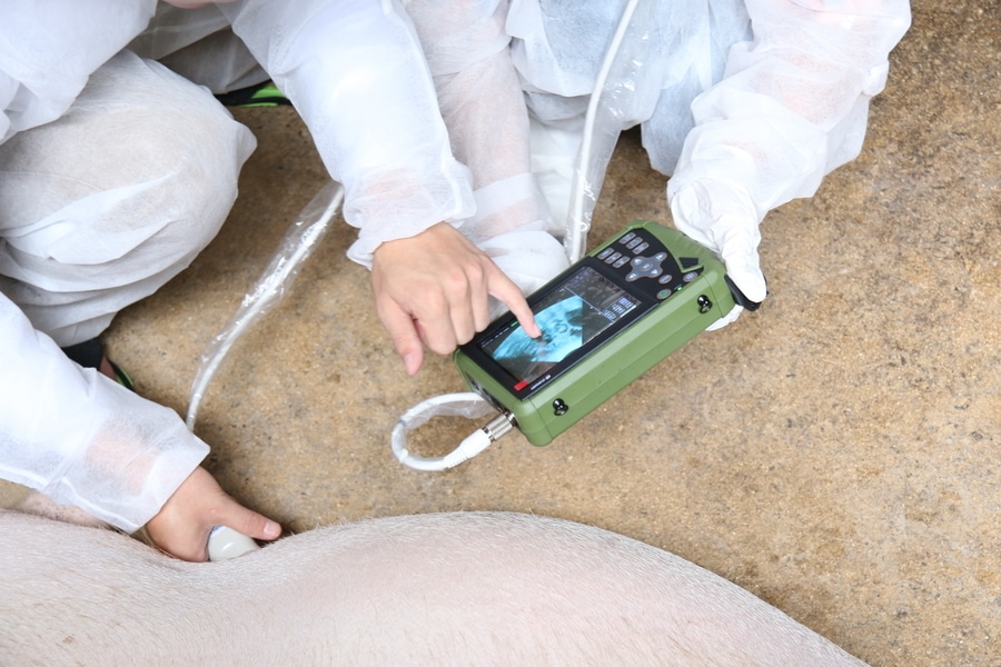
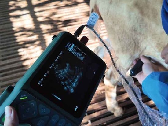
Ultrasound examination and Clinical Images of sows
Ultrasound Clinical Images of sows
Ultrasound images of sow pregnancy
The common veterinary ultrasound images generally show three colors: black, gray and white
1. Black: aqueous substances, such as amniotic fluid, blood, urine, pus, etc.
2. Gray: organ parenchyma.
3. white: denser objects, such as bones, teeth, organ fascia, etc.
A: 22-30 days after mating is the best stage to observe the image, showing a black irregular circle, it is clear and closer to the circle, and it’s easy to tell. At the same time, about 25 days is the best time to estimate the number of fetuses. One black circle represents a gestational sac, and will be a piglet in the future. The black circles in the left and right cornua uteri add up to the approximate number of babies conceived.
B: The image from 35 to 50 days after breeding is not very easy to tell, it shows a large black area looks like a big head and a small tail. If there are in large quantities, the entire display can be filled, some can see the white germ part inside the big black circle. Because of the development of the germ, the amniotic fluid is relatively less, so the black area of the image is not black enough compared to the early stage.
C: During the period from 60 days of breeding to farrowing, we mainly observe the skeletal images of the piglets, which show an arc-shaped series of white bright spots, which is the image of piglet spine. At the same time a ray-like image will appear on the display. An experienced user can observe the fetal heartbeat.
Possibility of misdiagnosis when using veterinary ultrasound
1、The user is not careful enough, not observing the image carefully, pursuing fast speed and not judging the image carefully.
2、The sow's belly is too dirty and the probe does not fully touch the skin, resulting in unclear images that affect the user's judgment.
3、The sow's bladder accumulated too much urine and the bladder swelled to obscure the uterine horn, which only showed a big black circle on the image.
4、The ultrasound machine is too low-end, the image quality is poor, and the user cannot judge normally.
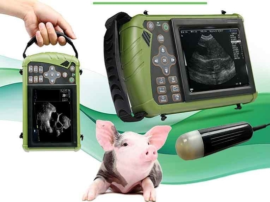
Dawei Veterinary Medical has professional technicians to guide you in using the veterinary ultrasound machine and provide you with comprehensive and perfect after-sales service.


