#Industry News
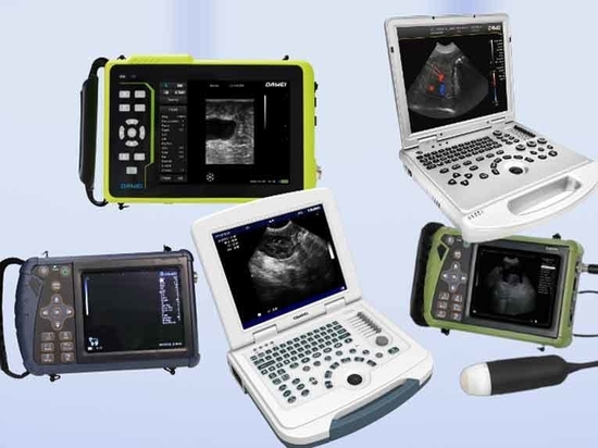
How to Use Dawei Veterinary Ultrasound Machine
How to Use Dawei Veterinary Ultrasound Machine
Veterinary ultrasound is an essential tool in modern livestock farming and pet diagnosis. Using ultrasound imaging technology, it enables non-invasive examination of internal animal structures and is widely used in pregnancy diagnosis, disease screening, and reproductive management. Proper operation not only improves diagnostic efficiency but also extends the lifespan of the equipment.
Preparation for Ultrasound Examination
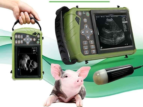
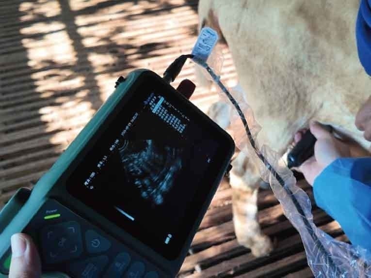
Before using the veterinary ultrasound machine, both equipment setup and animal preparation must be completed. First, check whether the main unit, probe, power cord, and other components are intact. Ensure proper grounding of the device to avoid electric shock risks. Choose the appropriate probe type and frequency based on the animal's size. Large animals such as cattle and horses require a rectal probe, while small animals like dogs and cats can be examined using an abdominal probe. For cattle and horses, a medium to low-frequency probe (3.5-5MHz) is recommended to balance penetration depth and resolution, while for pets, a high-frequency probe (above 7.5MHz) provides better image details.
For animal preparation, shave the examination area (such as the abdomen or rectal region) to remove hair and dirt, preventing interference with ultrasound transmission. Apply a sufficient amount of medical-grade coupling gel to eliminate air gaps between the probe and skin, ensuring effective sound wave penetration. For large animals like cattle and horses, proper restraint is necessary before operation. Experienced personnel should assist with rectal examinations to prevent the animal from struggling, which could damage the probe or blur the image.
Operational Procedure
Once the ultrasound machine is turned on, follow these standardized steps:
Parameter Adjustment: Set the overall gain, time gain compensation (TGC), and image depth according to the examination target. For pregnancy diagnosis, adjust the focal position to the uterine area and increase gain appropriately for a clear view of the embryo.
Probe Handling: Hold the probe firmly, applying light pressure while moving it slowly and keeping it perpendicular to the animal’s body. For rectal examinations in large animals, insert the probe carefully to avoid excessive force that could damage the intestinal walls. If image noise or gaps appear, adjust the probe angle or reapply coupling gel.
Image Interpretation: In pregnancy diagnosis, look for gestational sacs, fetal heartbeat, and embryo count. For disease screening, compare echogenic differences between normal and abnormal tissues. For example, cysts appear as anechoic (dark) areas, while tumors may present as mixed echogenic masses.
During operation, take the following precautions:
Never plug or unplug the probe while the machine is on to prevent circuit damage.
If the device emits abnormal noise or experiences image lag, turn it off immediately and contact technical support.
Important Notes
Operators must receive professional training. Unauthorized use by untrained personnel may cause animal distress and pose safety risks.
Avoid prolonged scanning. A single scan on the same area should not exceed 10 minutes to prevent potential tissue damage from ultrasound heat effects.
Do not bend or pull the probe cable. When storing, coil it into a loop with a diameter greater than 15 cm to prevent internal wire breakage.