#Livestock
The Role of Ultrasound Examination in Equine Lameness Cases
The Role of Ultrasound Examination in Equine Lameness Cases
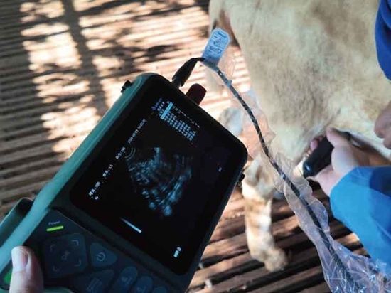
Ultrasound examination has become one of the most common on-site imaging modalities for evaluating musculoskeletal injuries in horses because it allows veterinarians to visualize almost any body tissue, most importantly soft tissues like tendons and ligaments. Ultrasound uses high-frequency sound waves to generate real-time images. The user places a probe that emits sound waves on the skin, directed towards the structure to be evaluated. When the sound waves encounter a structure or the interface between structures, they reflect back to the probe, much like sonar on a ship. The steeper the interface or the denser the structure, the more waves are reflected. The more sound waves received, the brighter the structure appears on the screen. We describe this brightness as echogenicity. For example, bones appear bright (echogenic), normal fluid is dark (anechoic), and all other structures are somewhere in between.
In cases of equine lameness, veterinarians are most likely to use ultrasound to assess tendons and ligaments, bone surfaces, synovial fluid, and cartilage. Tendons and ligaments can be imagined as ropes made of many strands or fibers. Tendons connect muscles to bones, while ligaments connect bones to each other. When tendons or ligaments are strained, their fibers may tear. Veterinarians assess the size, echogenicity, and fiber pattern of tendons or ligaments to evaluate the extent of their damage. Typically, minor tendon or ligament injuries result in an increase in their size or cross-sectional area. In cases of severe injury, veterinarians might notice changes in echogenicity and fiber pattern.
Normally, the “echo texture” or pattern of tendons or ligaments is uniform (consistently the same); a normal tendon’s transverse view shows a round or oval structure with uniform shading. A damaged tendon may appear round and bright (normal fibers) with dark areas. Dark areas indicate fiber tears or gaps where sound waves do not reflect. Larger, central fiber tear areas are often referred to as core lesions.
When observing the same area longitudinally, using the probe along the length of the tendon or ligament, normally long linear fibers may appear shorter and discontinuous or may disappear completely. Abnormalities are not always this obvious; real damage can be subtle, such as fine dark linear streaks or slightly irregular edges.
While ultrasound cannot penetrate bones, veterinarians can use it to evaluate bone surfaces. Due to the high density of bone, it should appear as a bright, white, smooth line on the screen. Changes in the bone surface around tendon or ligament insertions, arthritic joints, fractures, or osteochondritis dissecans (OCD) lesions can cause these lines to appear disrupted or rough.
Evaluating synovial structures (joints, tendon sheaths, and bursae) is equally useful. Normal structures have a membrane that produces a small amount of lubricating, nutrient-rich fluid. Inflammation from tendinitis, arthritis, direct trauma, or any other type of irritation causes the membrane to produce excessive, poor-quality fluid, sometimes rich in cells and proteins. Assessing synovial fluid and membranes can provide insight into the severity of inflammation. Additionally, veterinarians can check for defects in joint cartilage caused by trauma or OCD.
Using ultrasound for diagnosis is almost as important as using it for treating and monitoring injuries. For instance, in the case of tendon or ligament tears, veterinarians can inject regenerative products like stem cells or platelet-rich plasma directly into the fiber tear area under ultrasound guidance. They insert the needle into the ultrasound beam so they can visually see the penetration depth and observe the treatment product entering the space. Veterinarians can treat other areas like the sacroiliac joints, thoracolumbar spine, and cervical facet joints with anti-inflammatory medications under ultrasound guidance. Without ultrasound, they would perform treatments blindly, possibly too far from the pain site to be effective. Ultrasound guidance also ensures the needle does not inadvertently pierce other structures.
After an injury or treatment, veterinarians perform follow-up clinical and ultrasound examinations to assess healing. They look for reductions in the cross-sectional area of tendon and ligament injuries, increased echogenicity, and improved fiber alignment. Improvements seen in ultrasound examinations and clinical assessments together guide recommendations for increasing the horse’s workload.
Today’s veterinary ultrasound machines are portable, versatile, and accurate, making equine ultrasound an incredibly useful tool. With it, veterinarians can image any tissue to determine diagnoses while helping owners save money and time. It also aids in guiding the placement of therapeutic agents and monitoring recovery. If your veterinarian recommends an ultrasound for your horse, understanding its uses, mechanisms, and limitations can help provide clarity throughout the process.


