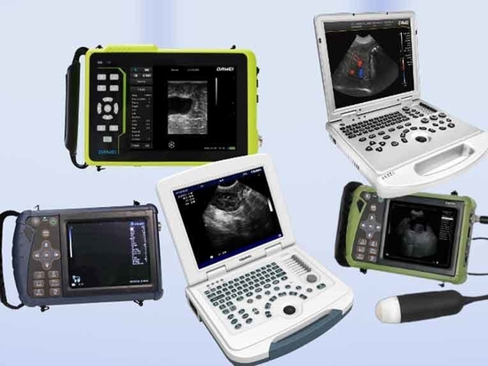
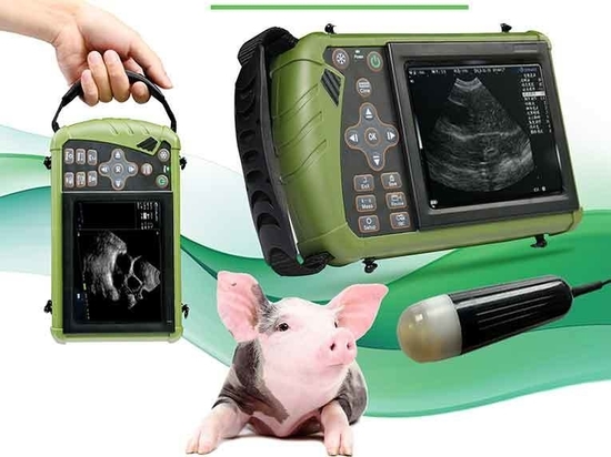
#Livestock
Camel ultrasonic machine application range and detection methods
Camel ultrasonic machine application range and detection methods
Camel from the previous heavy cargo consignment main function is slowly replaced by mechanical transportation equipment, however, has a rich nutritional value of camel milk more and more attention, camel milk powder contains very sufficient nutrients, including iron, calcium, protein, vitamin C, etc., quickly enhance immunity to strengthen the body. The broad market demand for camel milk continues to promote large-scale breeding of camels.
Camel ultrasound machine examination scope
Camel ultrasound working principle of other medical ultrasound working principle is basically the same, camel ultrasound machine working basic principle is through the ultrasound probe to the camel body to transmit a set of ultrasound waves, according to a certain direction for scanning. According to monitor the delay time of the echo and the strength of the echo pattern, you can determine the distance and nature of each organ, combined with pathology and clinical medicine, after observing, analyzing and summarizing a variety of reflective patterns, and then make a diagnosis of the site of the lesion, the nature of the dysfunction and the degree of a diagnosis.Abdominal organs: the liver, spleen, kidneys, gastrointestinal tract and other organs in the abdominal cavity can be examined.
Reproductive system: including ovaries, uterus, development of the fetus and so on. Using camel ultrasound veterinary ultrasound can expressly check whether the camel is successful in breeding and the number of fetuses whether there is stillbirth deformity heartbeat heart rate and other vital characteristics as well as quickly screen out female camels whether the uterus ovary inflammation hydrocele cysts and other diseases leading to camel does not come into heat and the cause of non-pregnancy, rapid enhancement of the camel reproduction and breeding and disease prevention ability, in the large-scale camel long is widely used.
Cardiovascular system: checking the structure and function of the heart, such as heart valves and heart muscle.
Respiratory system: to check the health of the lungs and airways.
Musculoskeletal system: to assess fractures, muscle injuries, etc.
Urinary system: including bladder, urethra, etc.
Subcutaneous tissue: detect abnormalities such as subcutaneous cysts, tumors, etc.
Camel ultrasound testing methods
Preparation: To ensure the propagation of ultrasound waves, first apply a conductive adhesive or lubricant to the surface of the camel’s skin. This helps to eliminate air gaps and improve image quality.
Probe selection: Choose the appropriate ultrasound probe according to the different parts of the examination. Generally, line array probes are used for superficial tissue examination and convex array probes are used for deep tissue examination.
Posture Adjustment: Depending on the examination site, the camel is allowed to adopt appropriate postures, such as standing, lying on its side, etc., in order to better expose the examination area.
Image Acquisition: The ultrasound probe is placed on the examination site and images are acquired at different angles by moving the probe. Ultrasound images are observed and recorded in real time for detailed evaluation of organs and tissues.
Image Analysis: The acquired ultrasound images are analyzed to identify abnormal structures or lesions. For example, the presence of liver lesions can be determined by observing the echo pattern of the liver, and heart valve function can be assessed by cardiac ultrasound.
Report Generation: A detailed examination report is generated based on the ultrasound results, including abnormalities found, recommendations for further examination needed, etc.
Ultrasound examination of camels requires professional veterinarians to operate and analyze to ensure accuracy and effectiveness.