#Livestock
Sheep pregnancy veterinary ultrasound
Sheep pregnancy veterinary ultrasound
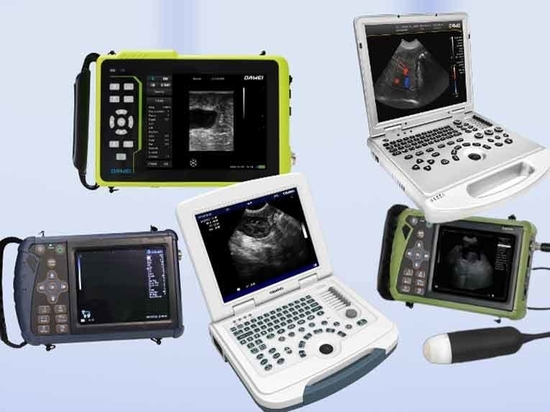
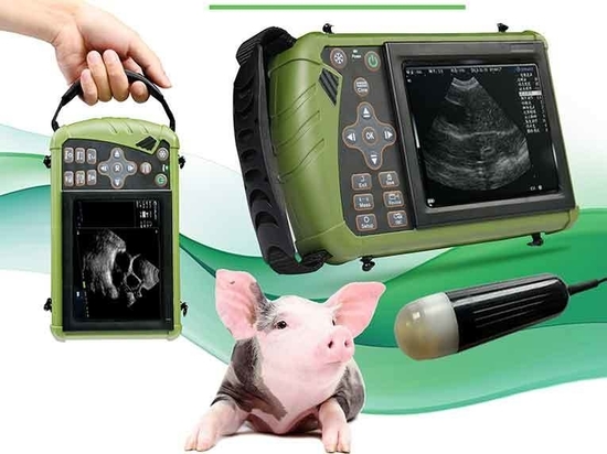
In the breeding industry, there have been more and more methods of pregnancy testing for animals, the most accurate of which is still diagnosed by ultrasound technology, commonly known as veterinary ultrasound machines, which are now widely used in pig and cattle breeding species. It is mainly used for fetal testing or to check for diseases of the uterus or ovaries.
The pregnancy cycle of sheep is about five months, ewes can be examined with veterinary ultrasound in more than thirty days of breeding, so how can ewes detect pregnancy? There are two ways to check ewes with ultrasound, one is an external abdominal ultrasound, the other is a rectal examination, both ways are more accurate, the former has the advantage of quickly checking whether there is a pregnancy or not, and the latter has the advantage of being able to distinguish more clearly through the rectal ultrasound whether there is a disease in the uterus or not.
In vitro examination, transabdominal large convex probe in vitro examination with a 3.5MHz convex array probe for surface examination, the best position is in the inner femur, the upper side of the back of the udder in the less hairy area. The ewe is lying on its side, the probe and the skin are coated with ultrasound coupling agent and then pressed vertically, and then moved closely to the skin at a uniform speed or at an appropriately altered angle, so that the ultrasound beam passes through the abdominal wall and the ultrasound cross-sectional echo images of the target monitoring tissues or organs are obtained.
Rectal examination, using sheep with handle 6.5MHz rectal probe into the rectum, the uterine horn, ovary sweep in turn, through the sweep, the screen will be rectangular picture to show the status of the body. 30~50 days after breeding is the best time to diagnose early pregnancy in sheep through rectal probe. 20~25 days of gestation in ewes, the fetal sac in the uterus can be detected at (8~12)×(12~16)mm and the fetal body; 25~30 days, the fetal heartbeat can be detected; after 30 days, the fetal sac, the fetal body, and the fetal heartbeat are very clear. 30~40 days, the fetal membranes can be detected; after 40~60 days, it is very easy to find out the fetal membrane; after 40~60 days, it is very easy to find out the fetal membrane; after 40~60 days, it is very easy to find out the fetal body. After 40 to 60 days of gestation, many cotyledonary structures can be easily detected, and the number of fetuses can be determined from the number of fetuses detected and the frequency of fetal heart beats by abdominal wall exploration in the ewe during 50 to 80 days of gestation.