#Livestock
Veterinary ultrasound diagnosis for pigs pregnancy
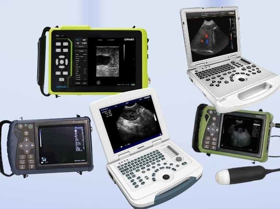
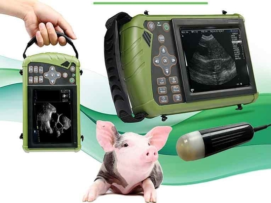
Pigs pregnancy ultrasound machine
With the use of veterinary ultrasound diagnostic technology, it is possible to detect whether a sow is pregnant or not about 20d after breeding. Dawei veterinary ultrasound equipment requirements, early pregnancy monitoring time, monitoring position, sow holding, monitoring methods and other methods are introduced as follows.
1 Terms and definitions
1.1 Veterinary ultrasound machine
Ultrasound is also known as ultrasound tomography. Veterinary ultrasound instrument is an ultrasound instrument dedicated to animal disease diagnosis, pregnancy diagnosis and livestock production. This use of large for veterinary SO ultrasound.
1.2 Ultrasound coupling agent
It is a medical product composed of water-based polymer gel. During ultrasonography, the air between the probe and the skin will prevent the ultrasound waves from being transmitted into the pig’s body, and in order to obtain high-quality and clear images, it is necessary to use a liquid conduction medium to connect the probe with the pig’s body surface, and this medium is the ultrasound coupling agent.
1.3 Time
The sows are mated from the 16th d to about the 35th d after breeding.
1.4 Body position
pig ultrasound
The posture presented by the sow’s body during ultrasound monitoring.
Detection range of ultrasound in contact with the skin during ultrasound monitoring at the acoustic window site
1.5 Sow holding
The control of the sow by means of manpower or instrumental equipment to limit the “defensive activity” of the sow.
1.6 Gain
Gain is an adjustment in the post-processing of ultrasound images due to the fact that ultrasound decreases in energy as it propagates. In order to compensate for this loss, the strength of echo energy can be artificially adjusted by post-processing to enhance the signal energy.
1.7 Contrast
The measurement of different brightness levels between the brightest white and darkest black in the image, the larger the range of difference represents the greater contrast, the smaller the range of difference represents the smaller contrast.
1.8 Glow
It is the subjective perception of the intensity of (achromatic) luminosity, or “a visual attribute that corresponds to the amount of light emitted from an area”. Glow indicates the intensity of light radiated from a surface.
2 Ultrasound monitoring operation points and techniques
Ultrasonic detection of pig
2.1 Monitoring time
Early pregnancy monitoring can be carried out 20d after sows are bred, and the monitoring time is 5d. Although the pregnancy sac can be detected on the 18th day after breeding, the detection rate is not high due to the small size of the pregnancy sac which is difficult to be found. On the 22nd to 24th day after breeding, the detection rate is extremely high, and on the 24th day the detection rate can reach 100%. In order to reduce human-caused pig emergencies, ultrasound detection can be carried out on the 22nd day.
2.2 Sow holding and body position
Sows can stand freely in the restricted feeding pen or lie on their sides in the holding pen for holding. Sow position is best in side-lying, crawling, standing or feeding, keeping quiet.
2.3 Sound window part
The sound window is usually located in the lower abdomen, the abdomen on the inside of the hind limb femur or the penultimate to the fourth pair of nipples. When the sows are in different physical conditions and in the state of full or empty stomach, the images are presented in different positions, and the scope of the sound window can be expanded to the periphery appropriately.
2.4 Dawei veterinary ultrasound machine S0 debugging
Switch on the ultrasound instrument and adjust the contrast, brightness and gain to suit the light intensity and the vision of the inspector.
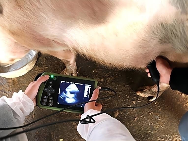
2.5 Monitoring operation
After determining the site of the sound window, subtract the abdominal hair of the pig, remove mud or dirt, and keep the probing site clean. The ultrasound probe was coated with ultrasound coupling agent and placed in the acoustic window site, so that the emitting surface of the ultrasound probe was closely connected with the skin, and the front and back up and down positions of the probe and the angle of incidence were adjusted. The probe was placed parallel to the body axis towards the urogenital tract of the sow for a sliding sweep or fan sweep, and after the bladder was detected, it was swept towards the upper or lateral part of the bladder.
2.6 Gestation determination
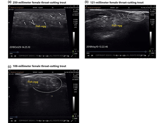
Positive pregnancy is diagnosed by showing a tomographic image of the blastocyst and embryo, i.e. the typical dark area of the gestational sac. The judgement of negative early pregnancy must be careful, the absence of a gestational sac is not equal to the absence of conception, may be due to the small number of conception or unskilled existence of missed detection. The area on both sides should be increased and carefully detected, and the test can be repeated several times until day 25 of breeding. A sow can be considered to be falsely pregnant if it is still not negative for early pregnancy on the 25th day of breeding.
If you would like to learn more about animal products, please feel free to consult with us.